Trichilemmal cyst
Hello everyone, how are you all? Its been so many days and again so much gap came and i was busy for month. I hope you all are doing well and happy and healthy. All Glory to God✨.
Today i would like to tell about "Trichilemmal cyst" also called pilar cyst and how it is diagnosed on histopathology.
A case of 60 year old man came to the hospital with a cystic swelling in the scalp.
So excision biopsy was done and sent for histopathology.
The cyst while doing grossing pultaceous keratin material seen. The tissue went into tissue processing.
Histopathology
4x magnification objective lens
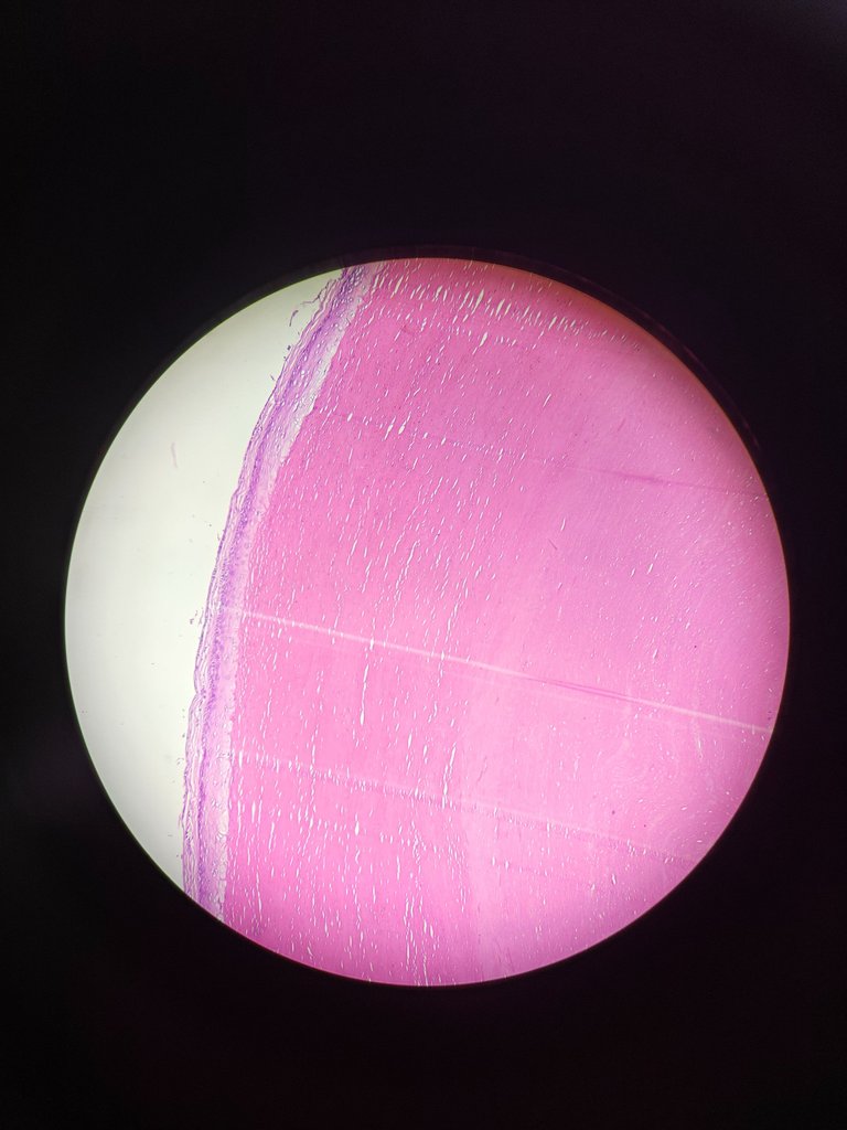
You can see in the above image shows cyst wall which is lined stratified squamous epithelium with absent granular layer. The cyst contains lamellated eosinophilic keratin.
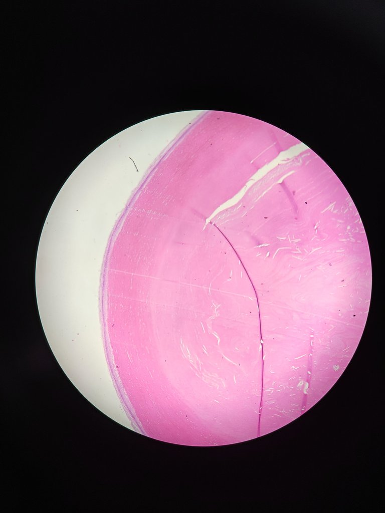
10x magnification of objective lens
 .
.
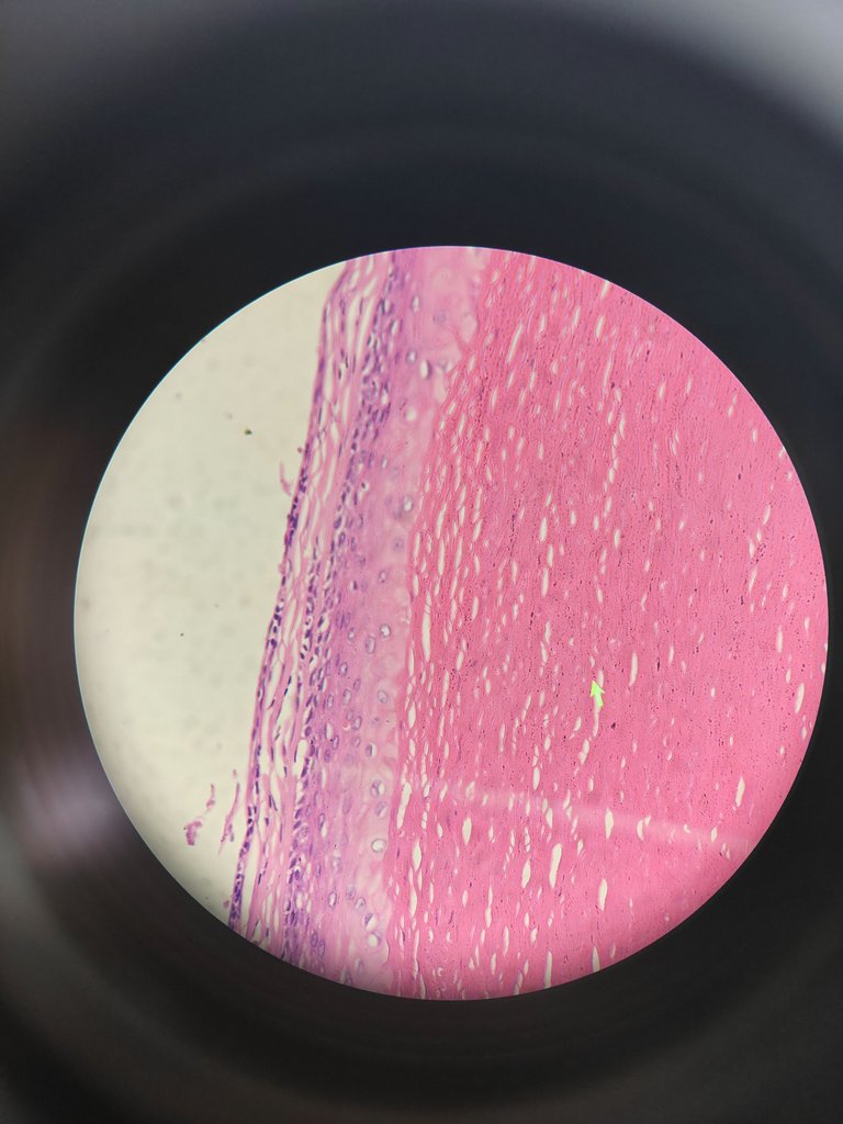
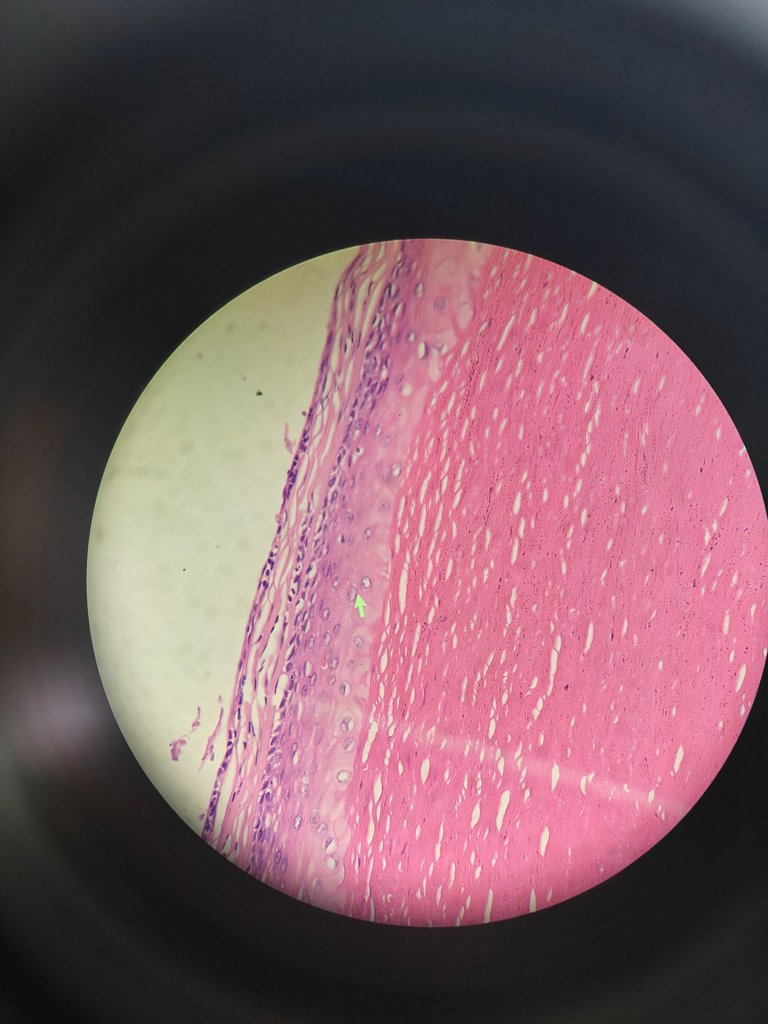
In the above image, the yellow cursor seen at foci
These are keratin flakes.
The below images have yellow color arrow pointing keratin flakes and stratified squamous epithelium
I hope you got it how trichilemmal cyst is diagnosed. I will come up with differential diagnosis for this entity in the next post. Keep learning and keep growing all.
References
- Lever's Dermatopathology: Histopathology of the Skin 12th Edition.
Thanks for reading,
with regards,
Thanks for your contribution to the STEMsocial community. Feel free to join us on discord to get to know the rest of us!
Please consider delegating to the @stemsocial account (85% of the curation rewards are returned).
Consider setting @stemsocial as a beneficiary of this post's rewards if you would like to support the community and contribute to its mission of promoting science and education on Hive.