CERVIX HISTOLOGY
Hello everyone😀, its been so long and i am back again. How are you all? Hope you all are doing well and me too. Today i would like to write on the topic of cervix histology.
First of all, what is cervix and where it is present?
Cervix is a part of female genital tract system and between isthmus of uterus and vagina it is present.
Why it is important to know about histology of cervix?
Cervical cancer is the 2nd most leading common cancer for females after breast cancer. This cervical cancer arises from cervix. So its always necessary to know what is normal histology of cervix.
Cervix is divided into two parts -
- Ectocervix
- Endocervix
Ectocervix is lined by stratified squamous epithelium
Endocervix is lined by simple columnar epithelium and has mucin in the lining epithelial cells.
We are going to see first ectocervix histology and how it looks like
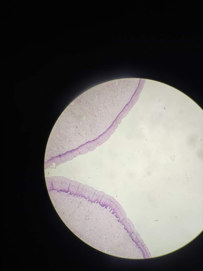
1. ECTOCERVIX
4X Magnification
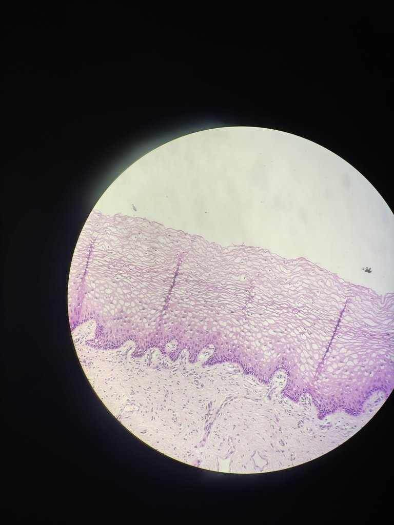
20X Magnification
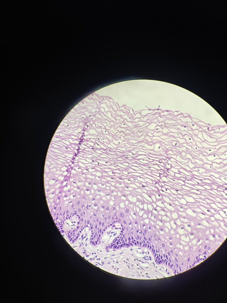
40X Magnification
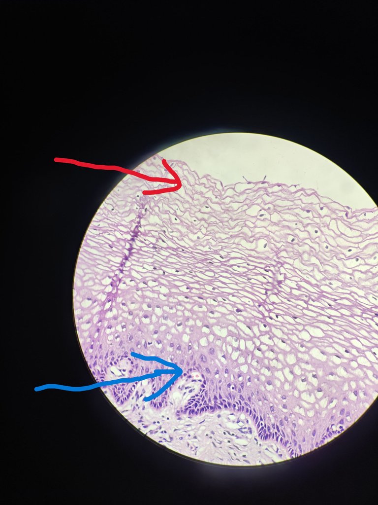
You can see in the above image that blue color arrow represents the basal layer and superficial layer is represented by red color arrow and there is loss nucleus in the superficial layer. The nucleus is preserved in the basal layer. So maturation happens when we go from basal layer to superficial layer and it normal. but if this maturation is not occuring in this way then there is some problem that we have to catch. The cytoplasm is clear because glycogen is present in it.
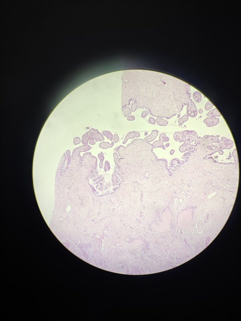
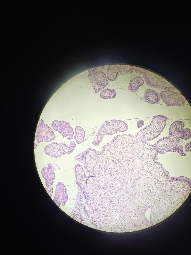
2.Endocervix
4X Magnification
10X Magnification
40X Magnification
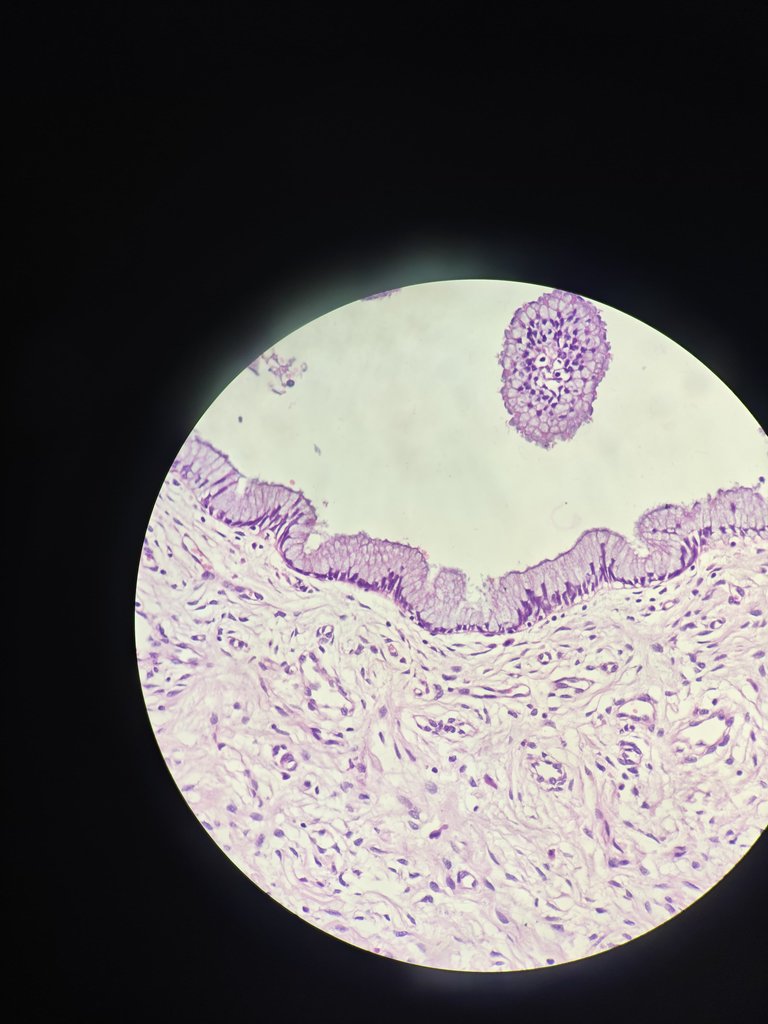
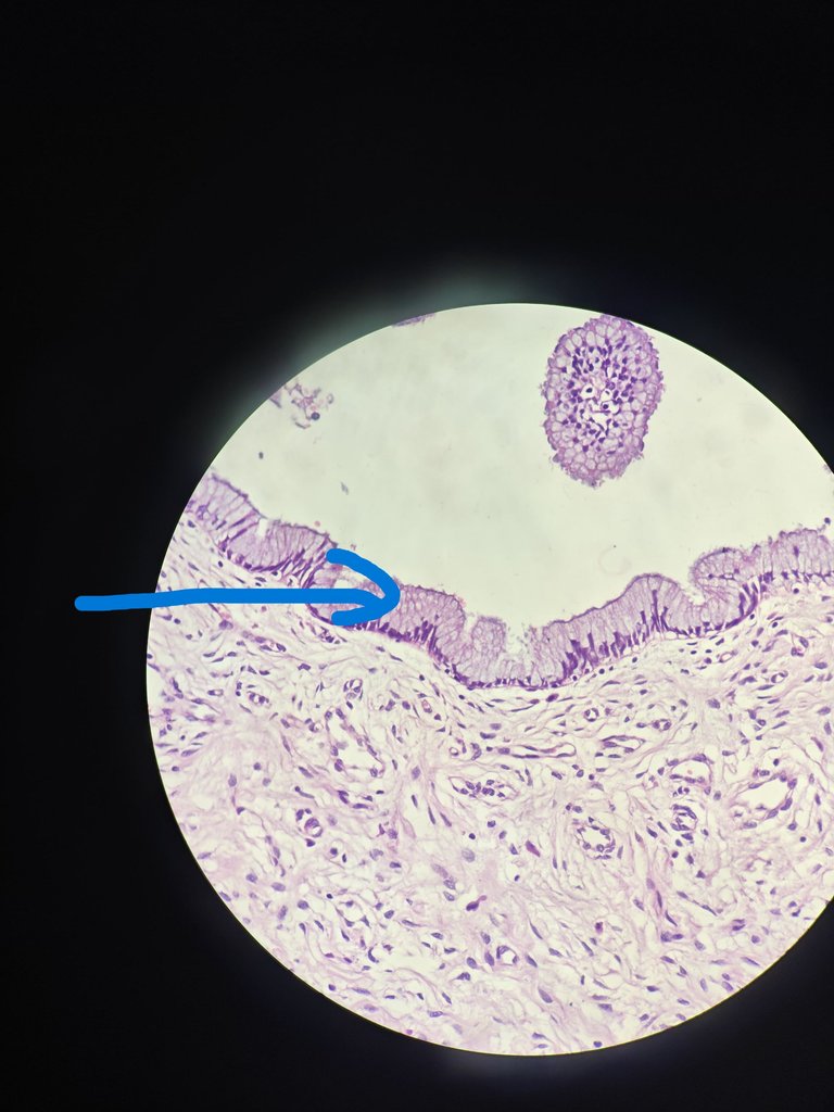
The Above image with blue arrow represents lining epithelium which is simple columnar epithelium with nucleus present at the base and cytoplasm has mucin.
I am going to share few images where it is junction of ectocervix and endocervix and you can see both lining epithelium and you will understand it. This can be called Transformational zone.
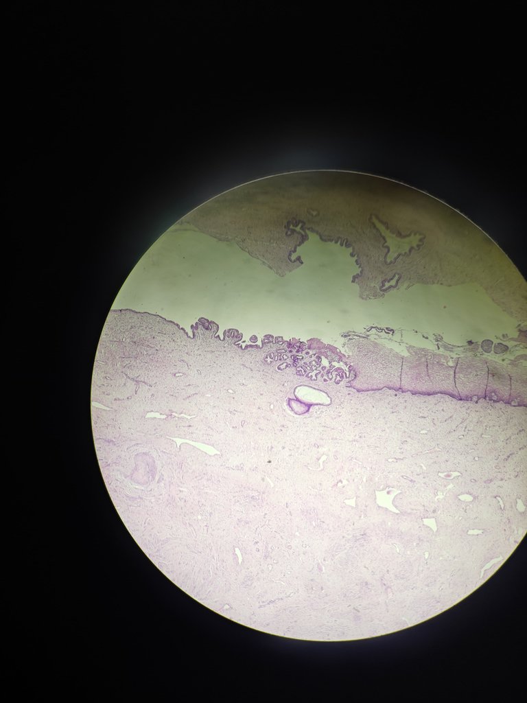
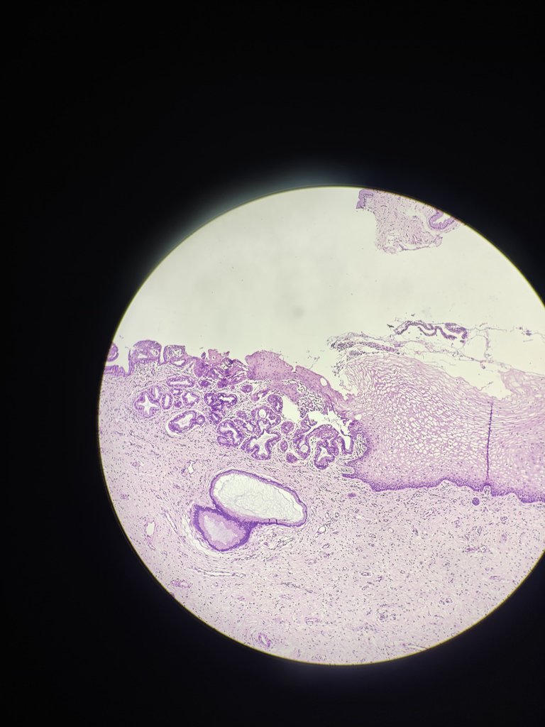
The left side of the image shows endocervix and right side of image shows ectocervix.
Squamous cell carcinoma arises from ectocervix and thats the reason PAP smear screening for females is very crucial to identify at early stage and manage it accordingly. even
Hope you understand histology of cervix.
References
- HISTOLOGY for PATHOLOGISTS FIFTH EDITION Stacey E. Mills, MD
Thanks for reading,
with regards
This post has been manually curated by @bhattg from Indiaunited community. Join us on our Discord Server.
Do you know that you can earn a passive income by delegating to @indiaunited. We share more than 100 % of the curation rewards with the delegators in the form of IUC tokens. HP delegators and IUC token holders also get upto 20% additional vote weight.
Here are some handy links for delegations: 100HP, 250HP, 500HP, 1000HP.
100% of the rewards from this comment goes to the curator for their manual curation efforts. Please encourage the curator @bhattg by upvoting this comment and support the community by voting the posts made by @indiaunited.
Thanks for your contribution to the STEMsocial community. Feel free to join us on discord to get to know the rest of us!
Please consider delegating to the @stemsocial account (85% of the curation rewards are returned).
You may also include @stemsocial as a beneficiary of the rewards of this post to get a stronger support.
I have seen histology slides in ages.
Nice photos.
Ohh thank you very much