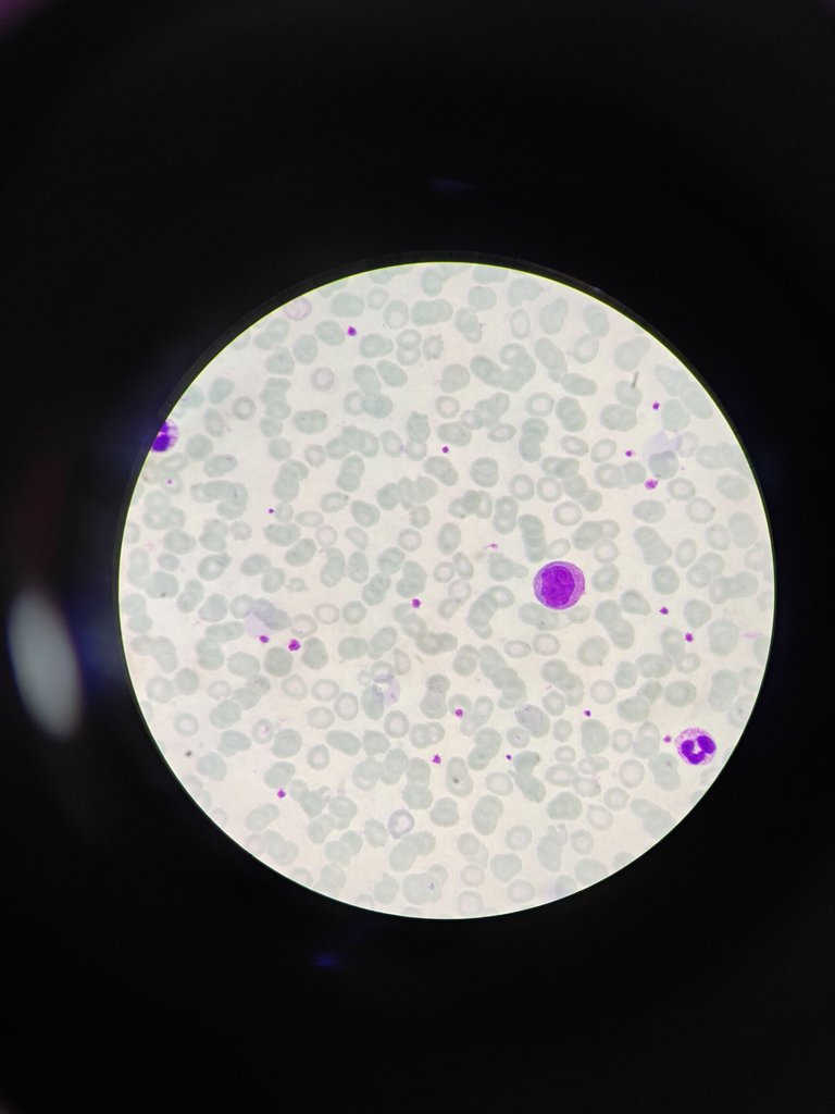
Cells in Peripheral smear
Hello everyone, how are you all? I hope you all are doing well and good and i am too. Now i haven't taken much gap to make a post again. So lets go into the post without wasting time.
Today i would like to share different kind of cells that we will see in a peripheral smear and these are present in our blood.
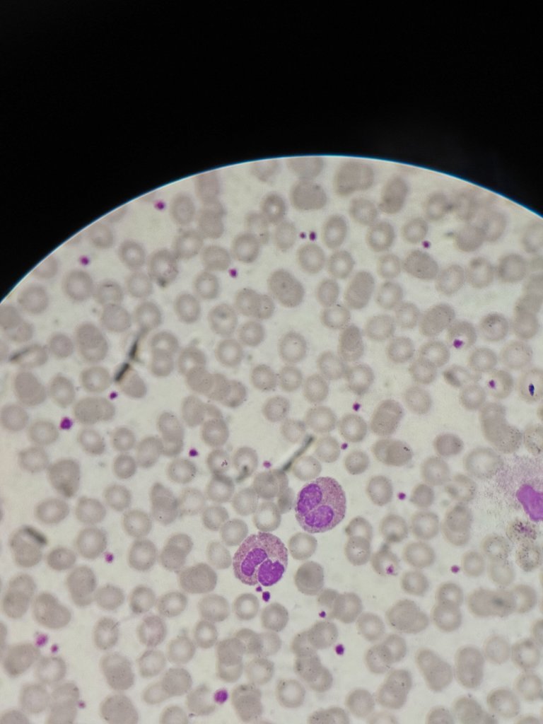
Neutrophils
These are more in blood compared to other cells and they are about 40-70% and they have multilobated nucleus about 3-5 lobes and in vit B12 deficiency they will become hyperlobated and that is one clue that patient is having vit 12 deficiency and cytoplasm has fine granules.
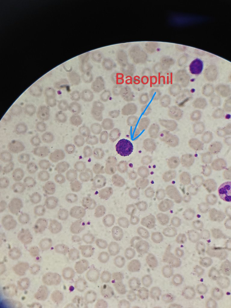
Basophil
These are least in number in peripheral blood infact you may not find them in peripheral smear also. They are about <1% only. They have bilobed nucleus and many basophilic granules are there and these granules helps in identifying them. They will increase in Chronic myeloid leukemia cases and it is called basophilia
Eosinophil
These are different kind of cells among all. They have bilobed nuclei and in literature it was given spectacle like nucleus and cytoplasm has eosinophilic granules which are numerous. They are about 1-6% only. But if there is increase in number of these cells then two things most commonly we have to remember that whether the patient is having allergy or parasitic infection.
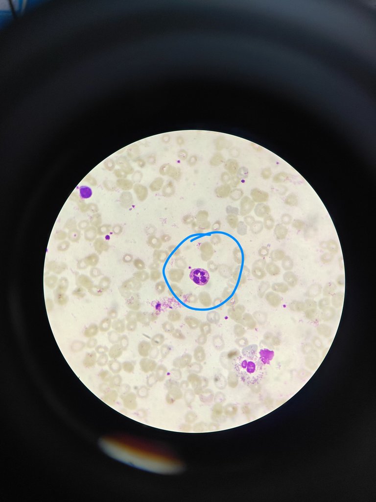
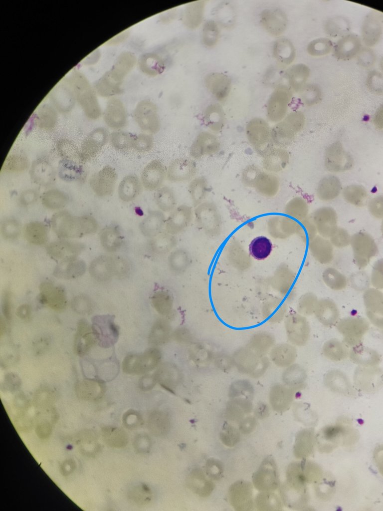
Lymphocyte
These are 2nd most commonest cell that we can see and they are small compared to other cells. This mature lymphocyte is has round nucleus which and cytoplasm is scant. They constitute about 20-40%. This cell is taken into consideration to classify RBC into microcytes and macroocytes the nucles of the lymphocyte is compared to size of RBC and also for lymphoma to call it as small cell or large cell lymphoma 2 lymphocyte size is taken as basic. These lymphocytes more newborns later they will reduce.
Monocyte
This is also largest cell like neutrophil. This constitutes about 8-10%. They have kidney shaped nucleus. And granules are seen. They will convert into macrophages in tissues helps in phagocytosis.
Hope you understand each cell and its significance.
Will come with new post.
References
- Dacie and Lewis Practical Haematology, Twelfth Edition,2017 Barbara J. Bain, Imelda Bates and Michael A. Laffan
- Wintrobe's Clinical Hematology, 14th Edition
Thanks for reading,
With regards,
Thanks for your contribution to the STEMsocial community. Feel free to join us on discord to get to know the rest of us!
Please consider delegating to the @stemsocial account (85% of the curation rewards are returned).
Consider setting @stemsocial as a beneficiary of this post's rewards if you would like to support the community and contribute to its mission of promoting science and education on Hive.